At GFN at UCM we have addressed the problem of high-resolution image reconstruction in PET for more than 10 years. We started collaborating with the Medical Image Lab of Hospital Gregorio Marañón in Madrid led by Dr. Manuel Desco. Our initial goal, in 2004, was to develop a software able to obtain high-quality images from PET acquisitions in a reasonable amount of time (i.e. less than 1 hour) from the acquired data in their scanners. Today, we have speeded-up the reconstruction more than 1000x respect to that initial goal, and we are able to reconstruct 3D high-quality images in a few seconds.
The use of accurate reconstruction methods is very challenging, as the number of variables to consider (voxels in the 3D images) and the amount of data measured, are both of the order of tens of Millions. Therefore, this big data problem required specific tools to handle it: simulation and compression of the system model, parallelization of the reconstruction algorithm, and the use of high-performance computers (clusters of computers and more recently, GPUs).
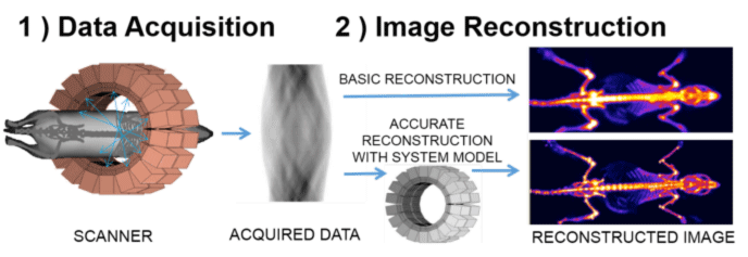
At GFN we proved that using all these tools it was possible to reconstruct high-quality PET images in just a few minutes (in 2004). The software that we developed, called FIRST, was registered and licensed to the Spanish company Suinsa S.A (Current Sedecal), for their preclinical PET scanner eXplore Vista, distributed worldwide for several years by General Electric, and later by other companies. The next figure shows an example of an image reconstructed with FIRST. This software is still in use in many prestigious research centers in the world such as Massachusetts General Hospital and Harvard Medical School, or John Hopkins University. I am currently working towards further reduction of the reconstruction time, with the ultimate goal of obtaining good-quality images in real-time during PET acquisitions. This would enable new applications, especially in surgical procedures.

Figure – Scheme of the Acquisition and Image Reconstruction Process. The reconstructed images were obtained with the standard method (right top) and with the software FIRST (right bottom) corresponding to a 18F-Na PET acquisition of a 200g rat using the eXplore Vista PET scanner. The improvement in resolution with FIRST is clear.
REFERENCES:
1. J.L. Herraiz, S. España, J.J. Vaquero, M. Desco, J.M. Udías. FIRST: Fast Iterative Reconstruction Software for PET Tomography. Physics in Medicine and Biology, 2006; 51: 4547-4565. PMID: 16953042.
2. J.L. Herraiz, S. España, E. Vicente, J.J. Vaquero, M. Desco, J.M. Udías. Noise and physical limits to maximum resolution of PET images. Nuclear Instruments and Meth. in Physics A, 2007.580(2):934-937.
DOI: 10.1016/j.nima.2007.06.039
3. J.L. Herraiz, S. España, R. Cabido, A. S. Montemayor, M. Desco, J. J. Vaquero, and J. M. Udias. GPU-Based Fast Iterative Reconstruction of Fully 3-D PET Sinograms. IEEE Transactions on Nuclear Science, 2011; vol. 58, no. 5: 2257-2263.
DOI: 10.1109/TNS.2011.2158113
|
